Our multinational team of trailblazing scientists explores nano-molecules with precision-crafted,
next-gen resolution, powered by state-of-the-art AI innovation.
| Group Members | |||
| Ali Shaib | Group Leader | ali.shaib(at)… | +49 (0) 551 39 65276 |
| Dr. Milton Mondal | Postdoc | milton.mondal(at)… | +49 (0) 55139 65994 |
| Donatus Krah | PhD Student | donatus.krah(at)… | +49 (0) 551 39 65740 |
| Antonios Ntolkeras | PhD Student | antonios.ntolkeras(at)… | +49 (0) 55139 65994 |
| Sushovan Chanda | PhD Student | sushovan.chanda(at)… | +49 (0) 551 39 65740 |
| Komal Rani | PhD Student | komal.rani(at)… | +49 (0) 551 39 65621 |
| Mohamad Mahdi Alawieh | Intern, Bachelor’s degree | mohamad.alawieh(at)… | +49 (0) 551 39 65621 |
| Alina Heimbrodt | Research Technical Assistant | alina.heimbrodt(at)… | +49 (0) 551 39 65892 |
| Jonas Altendorf | Master’s Student | jonas.altendorf(at)… | +49 (0) 551 39 65740 |
| Megi Pergjegjaj | Med, PhD Student | ||
| Maryam Baghai | Med, PhD Student | ||
About Us
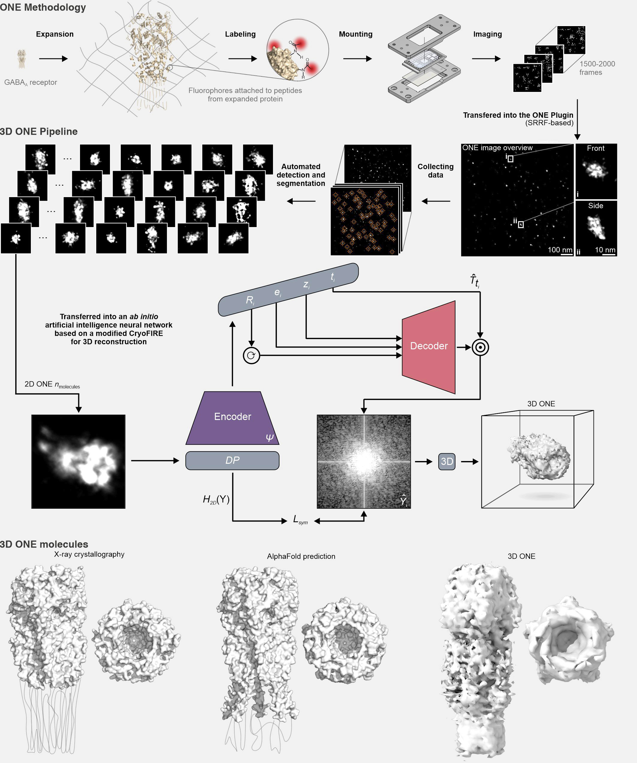
My group is based at the Department of Neuro- and Sensory Physiology, University Medical Center Göttingen, under the direction of Prof. Dr. Silvio O. Rizzoli. We developed One-step Nanoscale Expansion (ONE) microscopy, an innovative super-resolution technology that is transforming basic biological research. By combining the principles of expansion microscopy with fluorescence fluctuation analysis, this innovative method enables unparalleled visualization capabilities. It allows researchers to explore intricate details, from complex tissues to neuronal networks, and to delve into the ultra-structure of synapses. Furthermore, it extends to describing the shapes of individual protein molecules, bringing fluorescence microscopy to the threshold of “true molecular resolution” and unlocking unprecedented opportunities in structural biology.
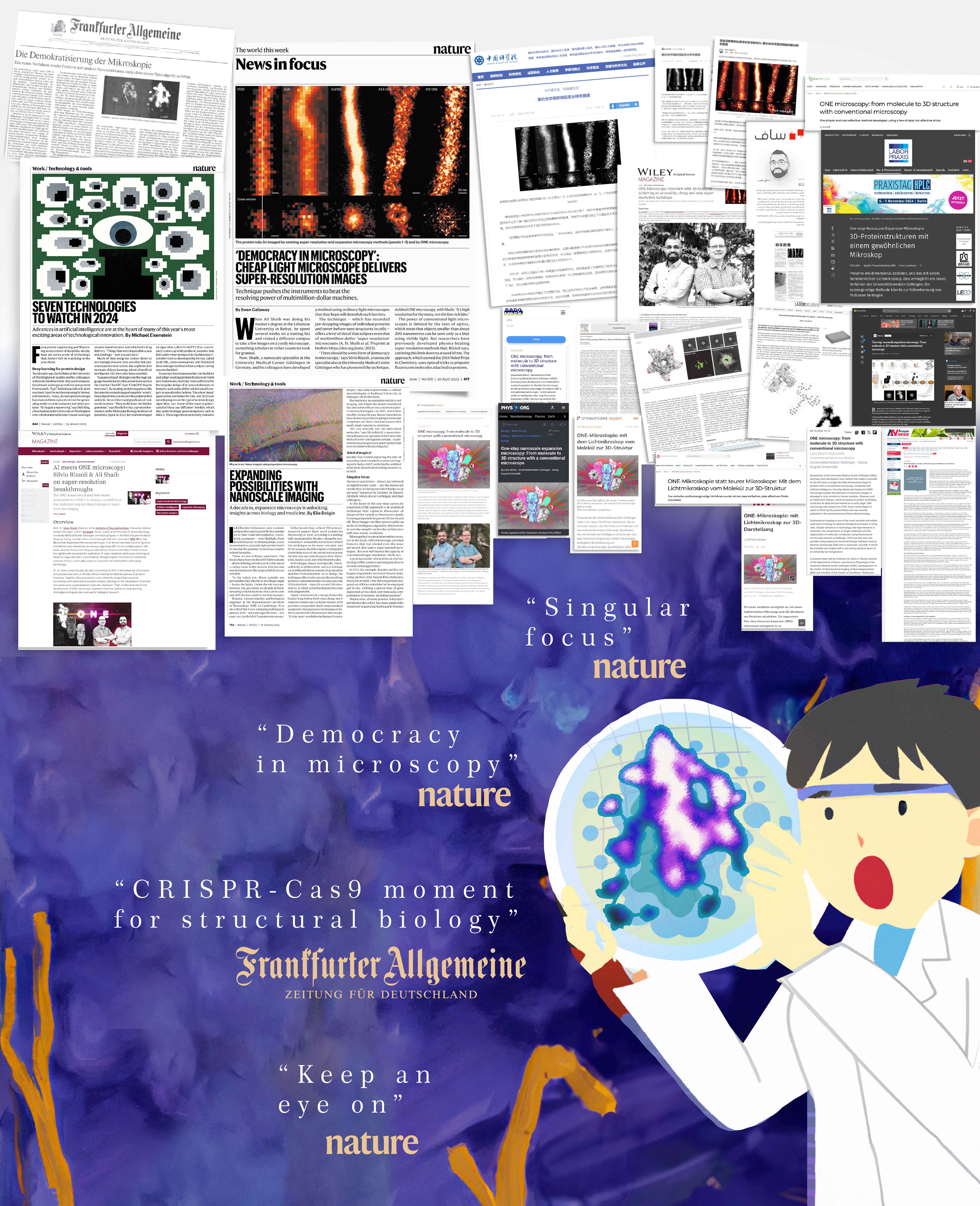
ONE microscopy achieves resolutions of 1 nanometer or better, granting labs equipped solely with fluorescence confocal systems access to a level of precision previously reserved for more specialized setups. This accessibility has been lauded in Nature as a milestone described as “democracy in microscopy,” a “singular focus,” and one of the “seven technologies to keep an eye on.” It has also been heralded by the Frankfurter Allgemeine Zeitung as a “CRISPR-Cas9 moment for structural biology,” reflecting its transformative impact on the field.
Using this technology, we have successfully expanded and imaged peptides, including mCLING, an eight-amino-acid peptide that labels membranes. Previously unresolvable by electron microscopy (EM) or X-ray crystallography due to its lack of density, mCLING can now be visualized in stunning detail. This breakthrough sheds light on previously inaccessible membrane-associated interactions, opening up new avenues of exploration.
Our research leverages this cutting-edge technology along with other non-fluorescence regimes like imaging mass spectrometry. These complementary approaches enhance our ability to explore complex biological systems and deepen our understanding of molecular and structural mechanisms. By providing valuable insights into these mechanisms, our work contributes to identifying molecular signatures important for disease detection. These studies are crucial for uncovering the cellular dysfunctions involved in conditions such as Parkinson’s disease, advancing both diagnostics and therapeutic development.
Artificial intelligence is revolutionizing our ability to analyze and interpret the vast data generated by ONE microscopy. Our dedicated team of AI specialists focuses on improving super-resolution capabilities and developing sophisticated pipelines for detection, segmentation, and 3D reconstruction of complex datasets. In addition, our basic research in AI is centered on computer vision techniques that incorporate specialized deep learning models for detecting rare events, such as vesicle fusion, synaptic transmission, and calcium waves, with improved accuracy and scalability. These advancements optimize the processing of fluorescence data, significantly reducing labor-intensive manual analysis and enhancing the precision and efficiency of biological investigations.
As a young and growing group, we are invested in addressing the challenges of large and complex biological datasets. We are also actively working to enhance the achievable resolution in imaging technologies, further pushing the boundaries of what can be visualized at the nanoscale. Especially in the AI segment, we aim to improve our ability to handle diverse datasets, develop reliable feature extraction methods, and create solutions for automating data analysis and visualization. Our focus is on refining these tools to support discoveries in fundamental research and explore potential applications in diagnostics and therapeutics.